Spirocerca lupi
Geographic Range
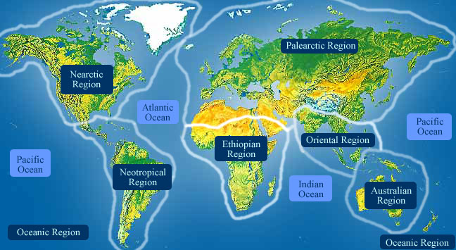
Spirocerca lupi, the esophageal worm, is a mammalian parasite found in the tropic warm temperate areas and throughout the United States. Their distribution is sporadic, as they have also been found in northern regions of the Soviet Union and Manchuria. (Frisby, 2001; Roberts and Janovy, 2000)
- Biogeographic Regions
- nearctic
- palearctic
- oriental
Habitat
Spirocera lupi most commonly plauge canids and wild felids in warm climates. However they have also been found in northern regions of the Soviet Union and Manchuria. (Roberts and Janovy, 2000)
- Terrestrial Biomes
- tundra
- taiga
- savanna or grassland
- chaparral
- forest
- rainforest
- scrub forest
Physical Description
Spirocerca lupi adults are cylindrical and range from a bright pinkish to red color. The mouth is sorrounded by six lips. Spirocerca lupi also have a well developed buccal capsule with thick walls. Females are approximately 5 to 8 cm long while males are slightly smaller, 3 to 5. 5 cm long. Cyndrical encapsulated juveniles that are passed out through the definitive host feces are 30-38 x 11-15 micrometers.
An outer cuticle has three main non-cellular outer layers made of collagen and other compounds that are secreted by the epidermis. The cuticle layer protects the nematodes so they can invade the digestive tracts of animals.
Nematodes have longitudinal muscles along the body wall. The muscles are obliquely arranged in bands. Dorsal, ventral and longitudinal nerve cords are connected to the main body of the muscle. (Brusca and Brusca, 2003; Roberts and Janovy, 2000)
- Other Physical Features
- ectothermic
- heterothermic
- bilateral symmetry
- Sexual Dimorphism
- female larger
- sexes shaped differently
-
- Range length
- 3 to 8 cm
- 1.18 to 3.15 in
Development
Embroynated S. lupi eggs exit the definitive host via feces. An intermediate host, a coprophagous (dung-eating) beetle, ingests the eggs while feeding. Inside the egg, the larva develops into an infective third stage and encyst in the intermediate host's tissues. The beetle is then eaten by a definitive host or by a paratenic host (lizard, chicken or mouse), which in turn is eaten by a definitive host. The third stage larva penetrates the stomach wall of the definitive host and migrates through the gastric wall and continues until it reaches the thoracic aorta within 20 days. It remains in the thoracic aorta for 2 to 3 months. After further development, they move to the esophagus where the larva becomes surrounded by a cystic nodule. At this point they can further develop and reach sexual maturity. (Frisby, 2001; Roberts and Janovy, 2000)
Reproduction
Adults are often found in clusters where mating occurs. Spirocera lupi are dioecious and after male and female find each other, via chemotactic an thigmotactic mechanisms, the males caudal papillae detects the female vulva. The male coils around a female with his curved area over the female genital pore. Males have copulatory spicules which they insert into the vulva. They do not conduct sperm, but hold the vulva open while the ejaculatory muscles inject sperm into her reproductive tract. Nematode sperm are amoeboid-like and lack flagella. (Brusca and Brusca, 2003; Pearse and Pearse, 1987; Roberts and Janovy, 2000)
- Key Reproductive Features
- sexual
- fertilization
- oviparous
- Parental Investment
-
pre-fertilization
- provisioning
Behavior
Spirocerca lupi migrate through their host by contracting and relaxing dorsal and ventral muscles. This forces the body into a series of curves producing an s-shaped motion. Adults are often found in clusters where mating occurs. Spirocera lupi are dioecious and after male and female find each other, via chemotactic an thigmotactic mechanisms, the males caudal papillae detects the female vulva. Males have copulatory spicules which they insert into the vulva. They do not conduct sperm, but hold the vulva open while the ejaculatory muscles inject sperm into her reproductive tract. (Pearse and Pearse, 1987; Roberts and Janovy, 2000)
Communication and Perception
Nematodes within the Secernentea have phasmids, which are unicellular glands. Phasmids likely function as chemoreceptors. Females may produce pheromones to attract males.
Nematodes in general have papillae, setae and amphids as the main sense organs. Setae detect motion (mechanoreceptors), while amphids detect chemicals (chemoreceptors). (Brusca and Brusca, 2003; Roberts and Janovy, 2000)
- Other Communication Modes
- pheromones
Food Habits
Parasitic nematodes feed on blood, tissue cells and fluid, intestinal contents or some combination of these. They also feed extravagantly with much waste. Spirocera lupi ingest food through their six lip mouth. A wave of muscle contractions pulls food into the digestive system. Pharyngeal glands and intestinal epithelium produce digestive enzymes to feed on the body fluids. Extracellular digestion begins within the lumen and is finished intracellularly. (Brusca and Brusca, 2003; Pearse and Pearse, 1987; Roberts and Janovy, 2000)
- Primary Diet
-
carnivore
- eats body fluids
- Animal Foods
- blood
- body fluids
Predation
These parasites are usually not preyed on directly, but are ingested from host to host. Larval mortality is high as most of the parasites do not reach appropriate hosts.
Ecosystem Roles
Spirocerca lupi eggs exit the definitive host (canine) via feces. An intermediate host, a coprophagous (dung-eating) beetle, ingests the eggs while feeding. Inside the egg, the larva develops into an infective third stage and encyst in the intermediate host's tissues. The beetle is then eaten by a definitive host or by a paratenic host (lizard, chicken or mouse), which in turn is eaten by a definitive host. (Frisby, 2001; Roberts and Janovy, 2000)
- Ecosystem Impact
- parasite
Economic Importance for Humans: Negative
Dogs infected with the esophageal worm can be severely damaged and even die. Nodules in an infected host can inhibit swallowing, breathing, and blood circulation. The dog may lose its appetite and weight or even have an aneurysm. Spirocerca lupi may also lead to the development of cancer and occasionally hypertrophic pulmonary osteopathy, inflamed and swollen joints. (Frisby, 2001; Roberts and Janovy, 2000)
- Negative Impacts
- causes or carries domestic animal disease
Other Comments
Signs of infection in dogs are vomiting, weight loss, and hemoptysis (coughing and spitting up blood). Eggs of S. lupi can also be found in feces or vomit. Endoscopic exams and x-rays can reveal the presence of nodules caused by the esophageal worm. Veterinarians treat infected animals with the drug disophenol. However, irreversible damage, such as a severe aneurysm or cancer cannot be treated effectively. (Frisby, 2001; Roberts and Janovy, 2000)
Contributors
Renee Sherman Mulcrone (editor).
Jemiah Cameron (author), University of Michigan-Ann Arbor, Teresa Friedrich (editor), University of Michigan-Ann Arbor.
Glossary
- Nearctic
-
living in the Nearctic biogeographic province, the northern part of the New World. This includes Greenland, the Canadian Arctic islands, and all of the North American as far south as the highlands of central Mexico.
- Palearctic
-
living in the northern part of the Old World. In otherwords, Europe and Asia and northern Africa.
- bilateral symmetry
-
having body symmetry such that the animal can be divided in one plane into two mirror-image halves. Animals with bilateral symmetry have dorsal and ventral sides, as well as anterior and posterior ends. Synapomorphy of the Bilateria.
- carnivore
-
an animal that mainly eats meat
- causes or carries domestic animal disease
-
either directly causes, or indirectly transmits, a disease to a domestic animal
- chaparral
-
Found in coastal areas between 30 and 40 degrees latitude, in areas with a Mediterranean climate. Vegetation is dominated by stands of dense, spiny shrubs with tough (hard or waxy) evergreen leaves. May be maintained by periodic fire. In South America it includes the scrub ecotone between forest and paramo.
- chemical
-
uses smells or other chemicals to communicate
- ectothermic
-
animals which must use heat acquired from the environment and behavioral adaptations to regulate body temperature
- fertilization
-
union of egg and spermatozoan
- forest
-
forest biomes are dominated by trees, otherwise forest biomes can vary widely in amount of precipitation and seasonality.
- heterothermic
-
having a body temperature that fluctuates with that of the immediate environment; having no mechanism or a poorly developed mechanism for regulating internal body temperature.
- internal fertilization
-
fertilization takes place within the female's body
- motile
-
having the capacity to move from one place to another.
- oriental
-
found in the oriental region of the world. In other words, India and southeast Asia.
- oviparous
-
reproduction in which eggs are released by the female; development of offspring occurs outside the mother's body.
- parasite
-
an organism that obtains nutrients from other organisms in a harmful way that doesn't cause immediate death
- pheromones
-
chemicals released into air or water that are detected by and responded to by other animals of the same species
- rainforest
-
rainforests, both temperate and tropical, are dominated by trees often forming a closed canopy with little light reaching the ground. Epiphytes and climbing plants are also abundant. Precipitation is typically not limiting, but may be somewhat seasonal.
- scrub forest
-
scrub forests develop in areas that experience dry seasons.
- sedentary
-
remains in the same area
- sexual
-
reproduction that includes combining the genetic contribution of two individuals, a male and a female
- tactile
-
uses touch to communicate
- taiga
-
Coniferous or boreal forest, located in a band across northern North America, Europe, and Asia. This terrestrial biome also occurs at high elevations. Long, cold winters and short, wet summers. Few species of trees are present; these are primarily conifers that grow in dense stands with little undergrowth. Some deciduous trees also may be present.
- temperate
-
that region of the Earth between 23.5 degrees North and 60 degrees North (between the Tropic of Cancer and the Arctic Circle) and between 23.5 degrees South and 60 degrees South (between the Tropic of Capricorn and the Antarctic Circle).
- tropical
-
the region of the earth that surrounds the equator, from 23.5 degrees north to 23.5 degrees south.
- tropical savanna and grassland
-
A terrestrial biome. Savannas are grasslands with scattered individual trees that do not form a closed canopy. Extensive savannas are found in parts of subtropical and tropical Africa and South America, and in Australia.
- savanna
-
A grassland with scattered trees or scattered clumps of trees, a type of community intermediate between grassland and forest. See also Tropical savanna and grassland biome.
- temperate grassland
-
A terrestrial biome found in temperate latitudes (>23.5° N or S latitude). Vegetation is made up mostly of grasses, the height and species diversity of which depend largely on the amount of moisture available. Fire and grazing are important in the long-term maintenance of grasslands.
- tundra
-
A terrestrial biome with low, shrubby or mat-like vegetation found at extremely high latitudes or elevations, near the limit of plant growth. Soils usually subject to permafrost. Plant diversity is typically low and the growing season is short.
References
Brusca, R., G. Brusca. 2003. Invertebrates. Sunderland, Massachusetts: Sinauer Associates, Inc..
Frisby, H. 2001. Spirocera Lupi. Foster and Smtih Pet Education, 81: 221-222.
Hamir, A. 1987. Esophageal perforation and phyothorax associatated with Spirocerca lupi infestation ina dog. The Veterinary Record, 19: 276.
Harris, S. 1996. Spirocerca lupi infection in the dog: abberant migration. The Journal of the American Animal Hospital Association, 32: 125-30.
Nolan, T. 2004. "Spirocerca lupi Homepage" (On-line). Diagnosis of Veterinary Endoparasitic Infections. Accessed September 28, 2004 at http://cal.vet.upenn.edu/dxendopar/parasitepages/filariidsandspirurids/s_lupi.html.
Pearse, V., J. Pearse. 1987. Living Invertebrae. Boston, Mass: Blackwell Publications.
Roberts, L., J. Janovy. 2000. Foundations of Parasitology. Boston: McGraw Hill.