Mansonella ozzardi
Geographic Range
Mansonella ozzardi is strictly a New World parasite that has never been found in the Old World. It was first studied in blood samples taken from Carib indians in British Guyana. The organism is located throughout northern Argentina, the Amazon drainage, the northern coast of South America, Central America and several islands of the West Indies. (Anderson, 1992; Roberts and Janovy, Jr., 2000)
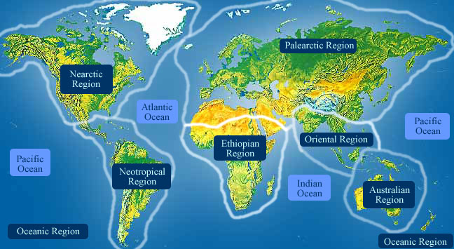
- Biogeographic Regions
- neotropical
Habitat
Mansonella ozzardi is concentrated in the tropical regions of the New World. Like all parasitic species, M. ozzardi requires nutrients from its hosts, and as an endoparasite it also requires the host to provide the habitat for it to survive. Adults live in the human body cavity among the mesenteries, peritoneum, and in the subcutaneous tissue, while juvenile stages require development in a Dipteran host. The immature worms migrate to the circulatory system of the human host, waiting to be ingested with an insect's blood meal. The factors that determine the suitability of particular Dipteran hosts has not yet been discovered, but after development is complete, the worms re-enter the human host when an infected insect bites another human. Thus, the entire life cycle of this organism is spent inside another species.
M. ozzardi may be especially well adapted to aboriginal Indians, in which the prevalence of infection is extremely high. In 1970, Marenhelle and German reported that 96.2% of adult Indians were infected in a region of Southern Colombia. (Anderson, 1992; Roberts and Janovy, Jr., 2000)
- Habitat Regions
- tropical
- Terrestrial Biomes
- forest
- rainforest
- scrub forest
- Other Habitat Features
- urban
- suburban
- agricultural
Physical Description
Mansonella ozzardi, being a nematode, is cylindrical, bilaterally symmetrical and possesses a pseudocoel, which is a fluid filled body cavity between the digestive tract and the body wall. The cuticle has three main outer non-cellular layers made of collagen and other compounds. The cuticle layer protects the nematodes so they can invade the digestive tracts of animals.
Nematodes have longitudinal muscles along the body wall. The muscles are obliquely arranged in bands. Dorsal, ventral and longitudinal nerve cords are connected to the main body of the muscle.
Adult Mansonella ozzardi are long and slender with reduced lips. Females are larger than males, and produce thousands of offspring called microfilariae, which are between 185-200 um long. Microfilariae of this species are unsheathed. When stained, the presence or absence of a sheath, internal nuclei and organs can all be seen, with the organization of these aiding in identification and classification of the different filarial worm species. (Anderson, 1992; Roberts and Janovy, Jr., 2000)
- Other Physical Features
- ectothermic
- heterothermic
- bilateral symmetry
- Sexual Dimorphism
- female larger
- sexes shaped differently
Development
Once in humans, the adults mate and females produce unsheathed microfilariae, which have a sharp tail. Females of this species are ovoviviparous, which is when embryos develop within the female's body, and the microfilariae produced are not as well differentiated as normal first stage juveniles. At this stage they are considered advanced embryos. The worms molt four times, the first two before hatching, and then before their adult stage. (Anderson, 1992; Bartoloni, et al., Nov., 1999; Roberts and Janovy, Jr., 2000)
Reproduction
Females may produce a phermomone to attract males. The male coils around a female with his curved area over the female genital pore. The gubernaculum, made of cuticle tissue, guides spicules which extend through the cloaca and anus. Males use spicules to hold the female during copulation. Nematode sperm are amoeboid-like and lack flagella. (Brusca and Brusca, 2003; Roberts and Janovy, Jr., 2000)
- Key Reproductive Features
- sexual
- fertilization
- ovoviviparous
- Parental Investment
-
pre-fertilization
- provisioning
-
pre-hatching/birth
-
provisioning
- female
-
provisioning
Behavior
The microfilariae wandering through the blood are ingested via a blood meal by species of biting midges of the genus Culicodes and black flies of the genus Simulium. The microfilariae are nonperiodic, thus they can be found in the peripheral circulatory system at all times. Upon entrance to the insect host, the microfilariae penetrate the body cavity and migrate to the thoracic muscles where development to infective stages takes place. In one study, the time frame in which the microfilariae in Culicoides furens migrated to the thorax was 24 hours. Once located in the thoracic muscles, M. ozzardi lays parallel to the muscle cells and over 2 to 3 days becomes shorter and fatter. After two molts, the larvae become active and migrate out from the thorax and go toward the head. Once here, they move down to the interior of the labium. When stimulated by warmth, the larvae escape the arthropod host when it takes a blood meal from a human. Back inside the human circulatory system, M. ozzardi makes its way to the internal body cavities and continues its growth and development to sexual maturity. (Anderson, 1992; Formica and Botto, 1990; Roberts and Janovy, Jr., 2000)
Communication and Perception
Nematodes within the Secernentea have phasmids, which are unicellular glands. Phasmids likely function as chemoreceptors. Females may produce pheromones to attract males.
Nematodes in general have papillae, setae and amphids as the main sense organs. Setae detect motion (mechanoreceptors), while amphids detect chemicals (chemoreceptors). (Brusca and Brusca, 2003; Roberts and Janovy, Jr., 2000)
- Other Communication Modes
- pheromones
Food Habits
Mansonella ozzardi is an endoparasitic organism that utilizes several Diptera species as an intermediate hosts and humans as a definitive host (the host in which sexually maturity is reached). Both entrance and exit from the definitive host occurs through the skin. Adults live in the human body cavity among the mesenteries, peritoneum, and in the subcutaneous layers feeding on the host's tissue fluid to grow. Infective larvae develop in the thoracic muscles of the arthropod host, once again feeding on the surrounding tissues to grow and molt.
Pharyngeal glands and intestinal epithelium produce digestive enzymes to feed on the hosts’ body fluids. Extracellular digestion begins within the lumen and is finished intracellularly. (Anderson, 1992; Brusca and Brusca, 2003; Roberts and Janovy, Jr., 2000; Yangco, et al., 1984)
- Primary Diet
-
carnivore
- eats body fluids
- Animal Foods
- body fluids
Predation
These parasites are probably not preyed on directly, but are ingested from host to host. Larval mortality is high as most of the parasites do not reach appropriate hosts.
Ecosystem Roles
Mansonella ozzardi is an endoparasitic organism that utilizes several species of Diptera as an intermediate host and humans as a definitive host. (Roberts and Janovy, Jr., 2000)
- Ecosystem Impact
- parasite
Economic Importance for Humans: Negative
The effects of Mansonella worms in a human can be profound but most infected hosts are asymptomatic. Increased age has been correlated with increased worm levels in the blood, which indicates that the most likely individuals to experience symptoms associated with M. ozzardi infection are those over the age of 25. Although most individuals are symptom-less, joint pains, headaches, coldness of the legs, and itchy red spots have been described in conjunction with M. ozzardi infection. Mansonella ozzardi is one of several filarial worm species associated with lymphatic filariasis. In addition, the manifestations of the parasite can mimic the more serious bancroftian filariasis, with polylymphadenitis, lymphedema, elephantiasis, and hepatomegaly over time.
The treatment of filarid infestations can vary. Conventional treatment has been with diethylcarbamazine (DEC) but recent studies have shown that Ivermectin can be an effective treatment of M. ozzardi depending on initial microfilariae levels. Also, prevention of the worms entering a human host relies heavily on insecticides and insect repellents. This action helps prevent infected Dipteran hosts from biting humans. (Bartoloni, et al., Nov., 1999; Gonzalez, et al., Dec., 1999; Orihel, et al., 1993; Roberts and Janovy, Jr., 2000)
- Negative Impacts
-
injures humans
- carries human disease
Contributors
Renee Sherman Mulcrone (editor).
Jason Prior (author), University of Michigan-Ann Arbor, Solomon David (editor), University of Michigan-Ann Arbor.
Glossary
- Neotropical
-
living in the southern part of the New World. In other words, Central and South America.
- agricultural
-
living in landscapes dominated by human agriculture.
- bilateral symmetry
-
having body symmetry such that the animal can be divided in one plane into two mirror-image halves. Animals with bilateral symmetry have dorsal and ventral sides, as well as anterior and posterior ends. Synapomorphy of the Bilateria.
- carnivore
-
an animal that mainly eats meat
- chemical
-
uses smells or other chemicals to communicate
- ectothermic
-
animals which must use heat acquired from the environment and behavioral adaptations to regulate body temperature
- fertilization
-
union of egg and spermatozoan
- forest
-
forest biomes are dominated by trees, otherwise forest biomes can vary widely in amount of precipitation and seasonality.
- heterothermic
-
having a body temperature that fluctuates with that of the immediate environment; having no mechanism or a poorly developed mechanism for regulating internal body temperature.
- internal fertilization
-
fertilization takes place within the female's body
- motile
-
having the capacity to move from one place to another.
- native range
-
the area in which the animal is naturally found, the region in which it is endemic.
- ovoviviparous
-
reproduction in which eggs develop within the maternal body without additional nourishment from the parent and hatch within the parent or immediately after laying.
- parasite
-
an organism that obtains nutrients from other organisms in a harmful way that doesn't cause immediate death
- pheromones
-
chemicals released into air or water that are detected by and responded to by other animals of the same species
- rainforest
-
rainforests, both temperate and tropical, are dominated by trees often forming a closed canopy with little light reaching the ground. Epiphytes and climbing plants are also abundant. Precipitation is typically not limiting, but may be somewhat seasonal.
- scrub forest
-
scrub forests develop in areas that experience dry seasons.
- sedentary
-
remains in the same area
- sexual
-
reproduction that includes combining the genetic contribution of two individuals, a male and a female
- suburban
-
living in residential areas on the outskirts of large cities or towns.
- tactile
-
uses touch to communicate
- tropical
-
the region of the earth that surrounds the equator, from 23.5 degrees north to 23.5 degrees south.
- urban
-
living in cities and large towns, landscapes dominated by human structures and activity.
References
Anderson, R. 1992. Nematode Parasites of Vertebrates; their development and transmission. Oxford: C.A.B. International.
Bartoloni, A., G. Cancrini, F. Bartalesi, M. Roselli. Nov., 1999. Mansonella ozzardi infection in Bolivia: Prevalence and clinical association in the Chaco region. Journal of Tropical Medicine & Hygiene, 61(5): 830-833.
Blaxter, M. 1996. "Mansonella ozzardi, Biology and Epidemiology" (On-line). Filarial Biology and Pathology. Accessed September 27, 2004 at http://nema.cap.ed.ac.uk/fgn/pnb/mansozz.html#path.
Brusca, R., G. Brusca. 2003. Invertebrates. Sunderland, Massachusetts: Sinauer Associates, Inc..
Formica, S., C. Botto. 1990. Filariasis focus due to Mansonella ozzardi and Mansonella perstans in Amazon Federal Territory of Venezuela. Journal of Tropical Medicine & Hygiene, 93(3): 160-165.
Gonzalez, A., D. Chadee, S. Rawlins. Dec., 1999. Ivermectin treatment of mansonellosis in Trinidad. West Indian Medical Journal, 48(4): 231-234.
Orihel, T., M. Eberhard, R. Lowrie. 1993. Mansonella ozzardi: The course of patency in experimentally infected patas monkeys. Tropical Medicine and Parasitology, 44(1): 49-54.
Roberts, L., J. Janovy, Jr.. 2000. Foundations of Parasitology sixth edition. United States: McGraw-Hill Companies.
Yangco, B., A. Vincent, A. Vickery, J. Nayar, D. Sauerman. 1984. A survey of filariasis among refugees in South Florida USA. American Journal of Tropical Medicine & Hygiene, 33(2): 246-251.