Gnathostoma spinigerum
Geographic Range
Although Gnathostoma spinigerum are considered endemic to Thailand, they are also found in many other countries of Southeast Asia. Reports of these nematode parasites have also been made in Japan, Australia, United States, and Mexico. The incidence of infection is more rare outside the Asian continent. (Daengsvang, et al., 1964)
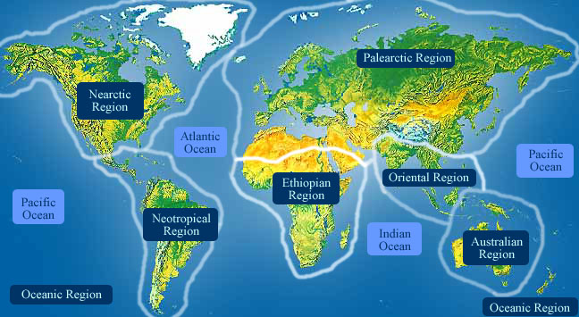
- Biogeographic Regions
- nearctic
- palearctic
- oriental
- neotropical
- australian
Habitat
Gnathostoma spinigerum can generally be found in wet tropical environments. Larvae can be found in many classes of animals that inhabit that ecosystem as well. While canine and feline species, and possibly pigs, are considered the definitive hosts, 36 naturally infected second intermediate hosts have been described along with additional experimentally determined potential hosts. In Japan, freshwater fishes, Ophicephalus argus and O. tadianus, are the most important vectors of human gnathostomiasis. In addition to freshwater fishes, domestic duck, Anas platyrhynchus and the domestic chicken Gallus gallus domesticus carry the parasite in Thailand. (Daengsvang, et al., 1966)
- Habitat Regions
- tropical
- freshwater
- Terrestrial Biomes
- forest
- rainforest
- Aquatic Biomes
- lakes and ponds
- rivers and streams
- Other Habitat Features
- urban
- suburban
- agricultural
Physical Description
Species within the genus Gnathostoma are characterized in part by a bulbous head with a pair of lateral lips surrounding a mouth on the longitudinal axis. The cephalic region is covered by transverse rows of cuticular spines. Internally, the head is divided into four glandular cervical sacs that attach near the esophagus, as well as four hollow spaces called ballonets, each being continuous with a cervical sac via a central canal. Despite having this structural knowledge, the functional aspects of these organs still remain a mystery. The body is typically pink and is also covered anteriorly with circles of flat, toothed spines, which become sparser and single-tipped further toward the end. After a bare region that constitutes roughly half of the body length, many small spines can be found on the posterior tip. Of the four species that can cause human gnathostomiasis--G. doloresi, G. hispidum, G. nipponicum, and G. spinigerum--differences in these external characteristics aid greatly in the identification of advanced third-stage larvae.
As a nematode, Gnathostoma spinigerum is cylindrical, has a cuticle with three main outer layers made of collagen and other compounds. The outer layers are non-cellular and are secreted by the epidermis. The cuticle layer protects the nematodes so they can invade the digestive tracts of animals.
Nematodes have longitudinal muscles along the body wall. The muscles are obliquely arranged in bands. Dorsal, ventral and longitudinal nerve cords are connected to the main body of the muscle.
As a nematode in the group Secernentea, Gnathostoma have specialized tubular excretory system with three canals. The canals are arranged to form an “H”.
On average, female worms are larger than males by an estimated 4 mm in length and 0.65 mm in width. Respectively, their size ranges from 11 to 54 mm and 11 to 31 mm long. Females are distinct in that they possess only two large papillae around their slightly rounded posterior ends. When viewed from the side, the dorsal outline is rounded, whereas the ventral side appears flat. Males have eight caudal papillae encompassing the anus on their bluntly rounded ends. Characteristic of males is the presence of blunt spicules that play the important reproductive role of holding open the vagina during sperm transfer. These male copulatory organs measure 1.1 mm and 0.4 mm long.
Just before molting into an adult, fourth-stage larvae have eight transverse rows of cephalic hooklets. At this point of development males can easily be differentiated from females based on identifiable sexual organs.
Advanced third-stage larvae bear four rows of hooklets on their head bulbs and measure 3 to 4 mm in length and 0.3 to 0.4 mm wide. The distinctive swollen head morphology, in addition to the four rows of hooklets, already becomes apparent in second-stage larvae.
Eggs have a polar cap at only one end and are laid unembryonated within pitted shells. Their dimensions are 65-70 µm by 38-40 µm. (Barnes, 1987; Maleewong, et al., 1995; Maleewong, et al., 1992; Roberts and Janovy, 2000; Vargas-Ocampo, et al., 1998)
- Other Physical Features
- ectothermic
- heterothermic
- bilateral symmetry
- Sexual Dimorphism
- female larger
- sexes shaped differently
-
- Range length
- 11 to 54 mm
- 0.43 to 2.13 in
Development
Eggs are released into the environment with the passing of feces by the canine or feline definitive host. At 27-31°C, eggs complete embryonation and free-swimming first-stage larvae hatch within one week. After being ingested by cyclopoid copepods, the larvae continue development into second- and early third-stage larvae in 7 to 10 days. A second intermediate host, generally a fish but may also be an amphibian or other vertebrate, consumes the infected crustacean and provides the muscle tissue in which the larvae molt and become advanced third-stage larvae. At this point, the G. spinigerum larvae are very infective to their definitive hosts, but they may simply roam throughout the tissues of a wrong host without reaching sexual maturity. With 36 different species as paratenic hosts the worms are able to have a widespread distribution. Paratenic hosts include crustaceans, freshwater fishes, amphibians, reptiles, birds, and mammals. It is not uncommon for animals to harbor numerous larvae in their body; one large king cobra was found to be infected with 1,020 larvae. The life cycle is completed when advanced third-stage larvae molt in a definitive host's tissue and finally develop into the adult stage in the stomach wall. Egg production begins about 100 days following infection.
Maleewong et al. (1992) found a seasonal variation in the prevalence, worm burden, and worm size in northeastern Thailand. Prevalence and worm burden are highest during the wet August-September sampling periods, while worms reached their maximum sizes during the following November-December months. A probable explanation suggests that larvae may suppress full maturity until the rainy seasons to ensure that the eggs in dog faeces have a mode to transportation to ponds or rivers.
Daengsvang et al. (1964, 1970) showed third-stage larvae penetrated the incised, scratched, and even intact skin of mice, rats, and cats. The larvae then migrated to the muscles where they became encysted. (Daengsvang, et al., 1964; Daengsvang, et al., 1970; Daengsvang, et al., 1966; Maleewong, et al., 1992; Roberts and Janovy, 2000)
Reproduction
Females may produce a phermomone to attract males. The male coils around a female with his curved area over the female genital pore. The gubernaculum, made of cuticle tissue, guides spicules which extend through the cloaca and anus. Males use spicules to hold the female during copulation. Nematode sperm are amoeboid-like and lack flagella. (Barnes, 1987; Roberts and Janovy, 2000)
- Key Reproductive Features
- sexual
- fertilization
- oviparous
- Parental Investment
-
pre-fertilization
- provisioning
Behavior
Gnathostoma spinigerum larvae tend to migrate unpredictably throughout the tissues of paratenic hosts. This may take them to superficial layers of the skin, as is usually the case in humans, or may lead them to more delicate areas such as an eye, the brain, or the spinal cord. Grave consequences can result from severe damage to the nervous system. If the worms remain near the cutaneous layer, they cause relatively minor damage, although they can exit the skin spontaneously. Such migration is termed creeping eruption or cutaneous larva migrans. Upon finding abscessed regions within the skin, the larvae may lie dormant or continue forward leaving red trails indicative of worm infection. (Roberts and Janovy, 2000)
Communication and Perception
Nematodes within the Secernentea have phasmids, which are unicellular glands that likely function as chemoreceptors. Females may produce pheromones to attract males.
Nematodes in general have papillae, setae and amphids as the main sense organs. Setae detect motion (mechanoreceptors), while amphids detect chemicals (chemoreceptors). (Barnes, 1987; Roberts and Janovy, 2000)
- Other Communication Modes
- pheromones
Food Habits
Pharyngeal glands and intestinal epithelium produce digestive enzymes to feed on the hosts’ body fluids. Extracellular digestion begins within the lumen and is finished intracellularly.
Following ingestion of infective advanced third-stage larvae, the definitive host's stomach wall provides the natural location for the embedding of adult Gnathostoma spinigerum in a tumorlike growth.
As larvae, these worms may migrate throughout the body feeding on tissues and affect other organs: liver, lung, kidney, orbit, larynx, and the central nervous system (CNS). They have also been found encysted in the muscle tissue of experimentally and naturally infected animals within one to two weeks. Studies of G. spinigerum infection in mice and rats via penetration of skin show that larvae can be found in the musculature of the neck, forelegs, chest, and back as well as in the skin. After 29 days, all of the larvae in the flesh are found to be encysted. In severe cases of human gnathostomiasis that affect the CNS, necrotic and hemorrhagic damage to cerebrum, brain stem, and spinal cord tissue can lead to death. (Barnes, 1987; Daengsvang, et al., 1970; Daengsvang, et al., 1966; Roberts and Janovy, 2000)
- Primary Diet
-
carnivore
- eats body fluids
- Animal Foods
- body fluids
Predation
These parasites are probably not preyed on directly, but are ingested from host to host. Larval mortality is high as most of the parasites do not reach appropriate hosts.
Ecosystem Roles
While canine and feline species, and possibly pigs, are considered the definitive hosts, 36 naturally infected second intermediate hosts have been described along with additional experimentally determined potential hosts. In Japan, freshwater fishes, Ophicephalus argus and O. tadianus, are the most important vectors of human gnathostomiasis. In addition to freshwater fishes, domestic duck, Anas platyrhynchus and the domestic chicken Gallus gallus domesticus carry the parasite in Thailand. (Roberts and Janovy, 2000)
- Ecosystem Impact
- parasite
- cats, Felidae
- canines, Canidae
- pigs, Suidae
- cyclopoid crustaceans, Cyclopoida
- humans, Homo sapiens
- Ophicephalus argus
- Ophicephalus tadianus
- Anas platyrhynchus
- Gallus gallus domesticus
Economic Importance for Humans: Negative
Human gnathostomiasis is a common parasitic disease of adults and older children throughout Southeast Asia that usually results from the ingestion of raw or undercooked meat containing third-stage larvae of G. spinigerum. Symptoms indicative of larval migration tend to consist in periodic swelling and superficial creeping eruption. However, in their erratic wandering, worms may sometimes find their way into the CNS in a minority of patients. Such invasions yield a variety of lesions: meningitis, encephalitis, radiculomyelitis, subarachnoid and parenchymatous hemorrhage, and local infarction. Patients with more serious damage involving the CNS can present with sudden onsets of headache and radicular pain prior to paralysis of the extremities and loss of bladder control.
Within weeks to months, half of patients infected with larvae demonstrate full recovery. The fatality rate of the remaining patients who suffer permanent neurologic sequelae ranges from 8-25%. Treatment of gnathostomiasis involves drug and hormone therapy that may include corticosteroids, albendazole, and ivermectin. Albendazole has been shown to facilitate the effective surgical removal of worms by causing their migration to the more easily accessible layers of dermis in humans. Chemotherapy has been ineffective against this disease. (Lowichik and Siegel, 1995; Nishamura and Hung, 1997)
- Negative Impacts
- injures humans
- causes or carries domestic animal disease
Other Comments
The survivability of infective third-stage larvae of Gnathostoma spinigerum has been examined in various experiments, which demonstrated that bare larvae could remain viable for nearly a day in 20 percent solutions of sodium chloride, citric acid, or acetic acid. They can naturally survive longer if they are encysted and surrounded by host tissue. Larvae in the flesh of raw fish can also survive submergence in lime juice and in fermented foods for 5 days. In addition, at a temperature of 4°C, they can live for one month. Encysted larvae 1 cm deep in raw fish are quickly killed within 5 minutes of treatment in boiling water. Prevention remains the most practical solution in reducing the incidence of human gnathostomiasis. In endemic areas with traditions involving the consumption of raw freshwater fish, care should be taken in that the fish is well frozen before preparation. If fish or poultry are to be cooked, they must be submitted to enough heat to ensure thorough doneness.
Effective diagnosis and treatment of infection depends greatly on accurate identification of the worm. In Japan, a skin test employing G. spinigerum antigens has been developed with success. (Daengsvang, et al., 1964; Roberts and Janovy, 2000)
Contributors
Renee Sherman Mulcrone (editor).
James Tseng (author), University of Michigan-Ann Arbor, Teresa Friedrich (editor), University of Michigan-Ann Arbor.
Glossary
- Australian
-
Living in Australia, New Zealand, Tasmania, New Guinea and associated islands.
- Nearctic
-
living in the Nearctic biogeographic province, the northern part of the New World. This includes Greenland, the Canadian Arctic islands, and all of the North American as far south as the highlands of central Mexico.
- Neotropical
-
living in the southern part of the New World. In other words, Central and South America.
- Palearctic
-
living in the northern part of the Old World. In otherwords, Europe and Asia and northern Africa.
- agricultural
-
living in landscapes dominated by human agriculture.
- bilateral symmetry
-
having body symmetry such that the animal can be divided in one plane into two mirror-image halves. Animals with bilateral symmetry have dorsal and ventral sides, as well as anterior and posterior ends. Synapomorphy of the Bilateria.
- carnivore
-
an animal that mainly eats meat
- causes disease in humans
-
an animal which directly causes disease in humans. For example, diseases caused by infection of filarial nematodes (elephantiasis and river blindness).
- causes or carries domestic animal disease
-
either directly causes, or indirectly transmits, a disease to a domestic animal
- chemical
-
uses smells or other chemicals to communicate
- ectothermic
-
animals which must use heat acquired from the environment and behavioral adaptations to regulate body temperature
- fertilization
-
union of egg and spermatozoan
- forest
-
forest biomes are dominated by trees, otherwise forest biomes can vary widely in amount of precipitation and seasonality.
- freshwater
-
mainly lives in water that is not salty.
- heterothermic
-
having a body temperature that fluctuates with that of the immediate environment; having no mechanism or a poorly developed mechanism for regulating internal body temperature.
- internal fertilization
-
fertilization takes place within the female's body
- marsh
-
marshes are wetland areas often dominated by grasses and reeds.
- motile
-
having the capacity to move from one place to another.
- native range
-
the area in which the animal is naturally found, the region in which it is endemic.
- oriental
-
found in the oriental region of the world. In other words, India and southeast Asia.
- oviparous
-
reproduction in which eggs are released by the female; development of offspring occurs outside the mother's body.
- parasite
-
an organism that obtains nutrients from other organisms in a harmful way that doesn't cause immediate death
- pheromones
-
chemicals released into air or water that are detected by and responded to by other animals of the same species
- rainforest
-
rainforests, both temperate and tropical, are dominated by trees often forming a closed canopy with little light reaching the ground. Epiphytes and climbing plants are also abundant. Precipitation is typically not limiting, but may be somewhat seasonal.
- sedentary
-
remains in the same area
- sexual
-
reproduction that includes combining the genetic contribution of two individuals, a male and a female
- suburban
-
living in residential areas on the outskirts of large cities or towns.
- swamp
-
a wetland area that may be permanently or intermittently covered in water, often dominated by woody vegetation.
- tactile
-
uses touch to communicate
- tropical
-
the region of the earth that surrounds the equator, from 23.5 degrees north to 23.5 degrees south.
- urban
-
living in cities and large towns, landscapes dominated by human structures and activity.
References
Barnes, R. 1987. Invertebrate Zoology. Orlando, Florida: Dryden Press.
Daengsvang, S., U. Chulalerk, T. Papasarathorn, B. Tongkoom. 1964. Epidemiological Observations On Gnathostoma spinigerum In Thailand. Journal of Tropical Medicine and Hygiene, 67: 144-147.
Daengsvang, S., B. Sermswatsri, P. Youngyi, D. Guname. 1970. Penetration Of The Skin By Gnathostoma spinigerum Larvae. Annals of Tropical Medicine and Parasitology, 64: 399-402.
Daengsvang, S., P. Thienprasitthi, P. Chomcherngpat. 1966. Further Investigations On Natural And Experimental Hosts Of Larvae Of Gnathostoma Spinigerum In Thailand. American Journal of Tropical Medicine and Hygiene, 15: 727-729.
Department of Parasitology, Faculty of Medicine, Chiang Mai University, THAILAND, 2004. "Nematode Image" (On-line). Department of Parasitology. Accessed 09/24/04 at http://www.medicine.cmu.ac.th/dept/parasite/nematode/framene.htm.
Lowichik, A., J. Siegel. 1995. Parasitic infections of the central nervous system in children. Part I: Congenital infections and meningoencephalitis.. Journal of Child Neurology, 10: 4-17.
Maleewong, W., P. Intapan, J. Khempila. 1995. Gnathostoma spinigerum: growth and development of third-stage larvae in vitro. The Journal of Parasitology v. 81 (Oct. '95) p. 800-3, 81: 800-803.
Maleewong, W., P. Loahabhan, C. Wongkham. 1992. Effects of albendazole on Gnathostoma spinigerum in mice. The Journal of Parasitology, 78: 125-126.
Nishamura, K., T. Hung. 1997. Current views on geographic distribution and modes of infection of neurohelminthic diseases. Journal of the neurological sciences, 145: 5-14.
Roberts, L., J. Janovy. 2000. Gerald D. Schmidt and Larry S. Roberts' Foundatoins of Parasitology, 6th edition. Boston: McGraw-Hill Higher Education.
Vargas-Ocampo, F., E. Alarcon-Rivera, F. Alvarado-Aleman. 1998. Human Gnathostomiasis in Mexico.. International Journal of Dermatology, 37: 441-444.