Capillaria hepatica
Geographic Range
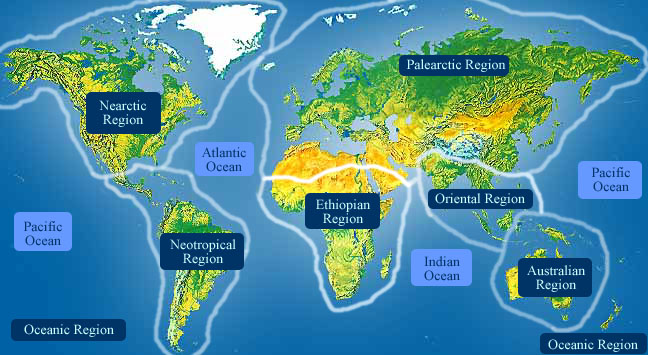
Capillaria hepatica has been recorded in Africa, Asia, Australia, Europe, and North, Central and South America. The eggs of C. hepatica can withstand various weather conditions and may remain unembryonated in the soil for long periods of time (> 1 year), which has contributed to the success of this species in several different climates. (Farhang-Azad, 1977; Li, et al., 2010; Olsen, 1986; Spratt and Singleton, 2001)
- Biogeographic Regions
- nearctic
- palearctic
- oriental
- ethiopian
- neotropical
- australian
Habitat
Capillaria hepatica is common in areas with poor hygiene and high rodent populations. On a smaller scale, within the body of its host, the larvae of C. hepatica hatch in the small intestine and spend most of their juvenile life migrating towards the liver, which is the final destination of the adults. The unembryonated eggs are excreted from the feces of the host and are then deposited in the soil. Ideal conditions for survival and embryonation of the ova are a cool and moist environment. (Conlogue, et al., 1979; Juncker-Voss, et al., 2000; Spratt and Singleton, 2001; Wright, 1974)
- Habitat Regions
- tropical
- terrestrial
- Terrestrial Biomes
- forest
- Other Habitat Features
- urban
- agricultural
- caves
Physical Description
This species can be identified as one of the two genera in the superfamily Trichuroidea that has bacillary bands, which consist of specialized cuticle and an underlying hypodermis extending the length of the body. Capillaria hepatica has cuticles shed at various points in the life cycle, characteristic of the nematodes. Adult males are 24-37 mm long and .04 to .08 mm in diameter. Adult females are larger than males and are approximately 53-78 mm in length and 0.19 mm in diameter. Capillaria hepatica has a muscular esophagus that is half of the females body and a third of the males body. The female's vulva is located posterior to the esophagus. The eggs are oval and in a double layer shell that has many minipores on it. The nonembryonated eggs are approximately 55.8 by 30 μm. (Conlogue, et al., 1979; Li, et al., 2010; Olsen, 1986; Spratt and Singleton, 2001; Tilney, et al., 2005; Wright, 1963)
- Other Physical Features
- ectothermic
- heterothermic
- bilateral symmetry
- Sexual Dimorphism
- female larger
-
- Average length
- 55 mm
- 2.17 in
Development
Capillaria hepatica is a special nematode because it is the only known helminth with a direct life cycle that requires the host to die for proper transmission. Eggs are deposited and embedded in the liver from the female worm and do not further develop until they are released from the organ. The eggs survive outside the host in an environment rich in oxygen, but nonetheless they are very resistant to the environment. They are liberated by cannibalism, predation, or necrophagy (feeding on carion) of an infected liver. After the new host consumes the egg, within 24 hours the egg hatches and the first stage larvae burrow into the cecal wall. The larvae move to the hepatic vein and the first molt in the larvae occurs approximately three days after the infection. Second stage larvae retain the shed cuticle as a sheath and a second molt takes place. Reproductive organs begin to form in the third larvae stage and the third molt takes place. The fourth stage larvae have a distinguished spicule and the cuticle is lost. This stage is also when sexual dimorphism is visible. Lastly, they will experience their final ecdysis, become fifth stage larvae and then adults. The female releases eggs in clumps around her body and then dies. The eggs become encapsulated in host tissue and protected until they begin development once released from the liver, upon death and decomposition of the host, or when the liver is eaten by a predator. If the liver disintegrates, the eggs are released into the soil and can be picked up by a new host. However, if cannibalism occurs, the eggs may pass through the predator undeveloped and be released in the feces. Then, they can embryonate and become infective for a new host because only embryonated eggs cause infection. (Bellows and Fisher, 1999; Farhang-Azad, 1977; Juncker-Voss, et al., 2000; Kumar, et al., 1985; Li, et al., 2010; Olsen, 1986)
Reproduction
Males have a long funnel-like spicule sheath that serves as a chemosensory organ. This likely serves in reproduction as the spicule helps hold the female during insemination. (Wright, 1974)
The spicule helps hold the female’s vagina during insemination. The females have a vulva located near the esophagus. Approximately twenty-eight days after mating, the production of eggs occurs. (Duggal and Kaur, 2007; Farhang-Azad, 1977; Juncker-Voss, et al., 2000; Olsen, 1986)
-
- Average number of offspring
- 457,783 eggs per liver
Females of Capillaria hepatica lay eggs in clusters surrounding the adults and the eggs become encapsulated and protected by host tissue. (Olsen, 1986; Spratt and Singleton, 2001)
- Parental Investment
- female parental care
- inherits maternal/paternal territory
Lifespan/Longevity
Laboratory trials found the lifespan of the female is about 59 days and about 40 days for males. (Li, et al., 2010)
-
- Range lifespan
Status: captivity - Females: about 59 days, Males: about 40 (high) days
- Range lifespan
Behavior
Capillaria hepatica is a non specific parasitic nematode that mostly affects rodents and rarely infects humans (there are less than 40 documented cases). The transmission of the eggs is unique because they remain in the liver until the host is eaten by a new host or until the host decomposes after death and embryonation takes place in the soil. Carnivorous animals living in a cliff habitat are the most frequently infected. The larvae of C. hepatica hatch in the small intestine and are then transported to the liver of the host where it lives as an adult. Eggs laid by the adult females become enclosed in host tissue and develop once released from the liver. (Conlogue, et al., 1979; Farhang-Azad, 1977; Spratt and Singleton, 2001)
Communication and Perception
Capillaria hepatica has cephalic sense organs that help in sensory perception. Their sensilla can function as chemoreceptors, in addition to mechanoreceptors. The centrioles in the sensory dendrites of C. hepatica consist of nine doublets. Additionally, the male’s sheath serves as a chemosensory organ to detect the female. (Wright, 1974)
Food Habits
Capillaria hepatica is surrounded by host liver cells and feeds from the enlarged cytoplasm of theses surrounding cells. This parasite is actually feeding from the reaction of the host rather than from blood or tissue fluid. Capillaria hepatica has thousands of bacillary pores that increase the surface area, as well as the capacity to absorb host nutrients. The bacillary pores are an advantage to living in the internal environment, such as the liver. (Wright, 1974)
- Primary Diet
-
carnivore
- eats body fluids
- Animal Foods
- body fluids
Predation
Capillaria hepatica is not preyed on directly. The eggs are durable in various environments; however, many eggs never reach a new host because release from the decaying liver or cannibalism is necessary for transmission. (Conlogue, et al., 1979; Li, et al., 2010; Spratt and Singleton, 2001; Tilney, et al., 2005; Wright, 1963)
Ecosystem Roles
Capillaria hepatica primarily lives in rodents and rarely infects humans. Once the eggs are ingested and arrive at the liver of the host, they cause serious damage to hepatic tissue. If released into the soil, the eggs may survive for extended amounts of time until embryonated (>1 year). (Farhang-Azad, 1977; Singleton and McCallum, 1990; Spratt and Singleton, 2001)
- Ecosystem Impact
- parasite
Economic Importance for Humans: Positive
Capillaria hepatica is an important biological control used especially in Australia to control the excessive rodent populations and the plagues that accompany them. Because transmission of Capillaria hepatica requires the death of the host, C. hepatica needs to be introduced early in the life cycle to see the desired result. If successful, there is a decrease in plague intensity and a very useful economic impact. C. hepatica is helping reduce the density of rodent pests in rural areas and improving agricultural crop survival. (Claveria, et al., 2005; Singleton and McCallum, 1990)
- Positive Impacts
- controls pest population
Economic Importance for Humans: Negative
Although there are economical benefits when the rodent population is infected, C. hepatica can be dangerous for humans. Capillaria hepatica causes a zoonotic disease called hepatic capillariasis, and rodents are the reservoir. Experiments have shown humoral immunity produced by mice after ingestion of C. hepatica eggs provide no protection against infection. Capillaria hepatica not only causes liver cell death, but additionally, it causes reduced fertility of the host. (Olsen, 1986; Spratt and Singleton, 2001)
- Negative Impacts
-
injures humans
- causes disease in humans
- carries human disease
Conservation Status
There is no effort to conserve or destroy this species. Although they can be used to control rodent populations, they still pose a pathogenic threat to humans.
-
- IUCN Red List
- Not Evaluated
-
- US Federal List
- No special status
-
- CITES
- No special status
-
- State of Michigan List
- No special status
Other Comments
Capillaria species are parasites in many vertebrate animals but only three species infect humans; Capillaria hepatica, Capillaria aerophila and Capillaria philippinensis. Also, the lesions on the liver can sometimes be mistaken for Schistosoma mansoni. (Govil and Desai, 1996; McCarthy and Moore, 2000)
Contributors
Hallie Leavitt (author), University of Michigan-Ann Arbor, Heidi Liere (editor), University of Michigan-Ann Arbor, John Marino (editor), University of Michigan-Ann Arbor, Barry OConnor (editor), University of Michigan-Ann Arbor, Renee Mulcrone (editor), Special Projects.
Glossary
- Australian
-
Living in Australia, New Zealand, Tasmania, New Guinea and associated islands.
- Ethiopian
-
living in sub-Saharan Africa (south of 30 degrees north) and Madagascar.
- Nearctic
-
living in the Nearctic biogeographic province, the northern part of the New World. This includes Greenland, the Canadian Arctic islands, and all of the North American as far south as the highlands of central Mexico.
- Neotropical
-
living in the southern part of the New World. In other words, Central and South America.
- Palearctic
-
living in the northern part of the Old World. In otherwords, Europe and Asia and northern Africa.
- agricultural
-
living in landscapes dominated by human agriculture.
- bilateral symmetry
-
having body symmetry such that the animal can be divided in one plane into two mirror-image halves. Animals with bilateral symmetry have dorsal and ventral sides, as well as anterior and posterior ends. Synapomorphy of the Bilateria.
- carnivore
-
an animal that mainly eats meat
- causes disease in humans
-
an animal which directly causes disease in humans. For example, diseases caused by infection of filarial nematodes (elephantiasis and river blindness).
- chemical
-
uses smells or other chemicals to communicate
- ectothermic
-
animals which must use heat acquired from the environment and behavioral adaptations to regulate body temperature
- female parental care
-
parental care is carried out by females
- forest
-
forest biomes are dominated by trees, otherwise forest biomes can vary widely in amount of precipitation and seasonality.
- heterothermic
-
having a body temperature that fluctuates with that of the immediate environment; having no mechanism or a poorly developed mechanism for regulating internal body temperature.
- motile
-
having the capacity to move from one place to another.
- oriental
-
found in the oriental region of the world. In other words, India and southeast Asia.
- oviparous
-
reproduction in which eggs are released by the female; development of offspring occurs outside the mother's body.
- parasite
-
an organism that obtains nutrients from other organisms in a harmful way that doesn't cause immediate death
- sexual
-
reproduction that includes combining the genetic contribution of two individuals, a male and a female
- tactile
-
uses touch to communicate
- terrestrial
-
Living on the ground.
- tropical
-
the region of the earth that surrounds the equator, from 23.5 degrees north to 23.5 degrees south.
- urban
-
living in cities and large towns, landscapes dominated by human structures and activity.
References
Bellows, T., T. Fisher. 1999. Handbook of biological control: principles and applications of biological. San Diego: Academic Press.
Camargo, L., J. Camargo, L. De Souza Vera, P. Barreto, E. Tourinho, M. de Souza. 2010. Capillariaisis (Trichurida, Trichinellidae, Capillaria hepatica) in the Brazilian Amazon: low pathogenicity, low infectivity and a novel mode of transmission. Parasites & Vectors, 3 (11): 1-6. Accessed April 16, 2011 at http://www.parasitesandvectors.com/content/3/1/11.
Claveria, F., J. Causapin, M. de Guzman, M. Toledo, C. Salibay. 2005. Parasite diversity in Rattus spp caught in wet markets. Southeast Asian J Trop Med Public Health, 36: Supplement 4.
Conlogue, G., W. Foreyt, M. Adess, H. Levine. 1979. Capillaria hepatica (Bancroft) in select rat populations of Hartford, Connecticut, with possible public health implications. Journal of Parasitology, 65 (1): 105-108.
Duggal, C., H. Kaur. 2007. Studies on the male and female copulatory apparatus of Trichuris globulosa (Nematoda, Trichuridae). Helminthologia, 44 (4): 151-156.
Farhang-Azad, A. 1977. Ecology of Capillaria hepatica (Bancroft 1893) (Nematoda). II. Egg-releasing mechanisms and transmission. The Journal of Parasitology, 63 (4): 701-706.
Govil, H., M. Desai. 1996. Capillaria hepatica parasitism. Indian Journal of Pediatrics, 63 (5): 698-700.
Juncker-Voss, M., H. Prosl, H. Lussy, U. Enzenberg, H. Auer, N. Nowotny. 2000. Serological detectionof Capillaria hepatica by indirect immunofluorescence assay. Journal of Clinical Microbiology, 38: 431-433. Accessed April 02, 2010 at http://jcm.highwire.org/cgi/content/full/38/1/431.
Kumar, V., J. Brandt, J. Mortelmans. 1985. Hepatic capillariasis may stimulate the syndrome of visceral larva migrans, an analysis. Annales de la Societe Belge de Medecine Tropicale, 65: 101-104. Accessed April 16, 2011 at http://lib.itg.be/open/ASBMT/1985/1985asbm0101.pdf.
Li, C., H. Yang, Y. Wang. 2010. Capillaria hepatica in China. World Journal Gastroenterology, 16: 698-702.
McCarthy, J., T. Moore. 2000. Emerging helminth zoonoses. International Journal for Parasitology, Volume 30: 1351-1360.
Olsen, O. 1986. Animal Parasites: Their Life Cycles and Ecology. New York City: Dover Publications.
Singleton, G., H. McCallum. 1990. The potential of Capillaria hepatica to control mouse plagues. Parasitology Today, 6 (6): 190-193.
Spratt, D., G. Singleton. 2001. Hepatic capillariasis. Pp. 365-367 in W Samuel, M Pybus, A Kocan, eds. Parasitic Diseases of Wild Mammals. Iowa: Iowa State University Press.
Tilney, L., P. Connelly, G. Guild, K. Vranich, D. Artis. 2005. Adaptation of a nematode parasite to living within the mammalian epithelium. Journal of Experimental Zoology Part A: Comparative Experimental Biology, 303A (11): 927-945. Accessed April 19, 2010 at http://www3.interscience.wiley.com/cgi-bin/fulltext/112101333/PDFSTART.
Wright, K. 1963. Cytology of the bacillary bands of the nematode Capillaria hepatica (Bancroft, 1893). Journal of Morphology, 112 (3): 233-259.
Wright, K. 1974. Cephalic sense organs of the parasitic nematode Capillaria hepatica (Bancroft, 1893). Canadian Journal of Zoology, 52: 1207–1213.